
Researchers at the Jagiellonian University have developed the world's first method for quickly recognizing bacteria and fungi in images from light microscopes. The new technology uses deep neural networks and artificial intelligence. Ultimately, it is expected to support the work of laboratory diagnosticians and doctors, but it will also be able to be used in industry, food safety monitoring or research work. The Krakow university wants to put the new solution into clinical use as soon as possible.
Neural networks and AI will speed up disease diagnosis. Researchers at the Jagiellonian University have developed an important tool

Published April 9, 2024 10:02

fot. UJ
- For the user of this solution, everything seems to be very simple. One has to send to the computer system images of microorganisms taken under the microscope. In response, the system generates a report with a list of specific species of bacteria or fungi that are present in the examined material. The whole operation takes no more than a minute," explains Bartosz Zieliński, Ph.D., professor at the Jagiellonian University's Institute of Computer Science and Mathematics.
Detect microorganisms in seconds
What seems simple on the part of the system operator is much more complex on the part of the technology required to run such a solution. And behind it are, among other things, deep neural networks and the nearly decade-long results of interdisciplinary research work carried out under the direction of Prof. Monika Brzychczy-Włoch, PhD, head of the Department of Molecular Medical Microbiology at the Jagiellonian University, and Bartosz Zielinski, PhD, Prof. of the Jagiellonian University, from the Institute of Computer Science and Computer Mathematics at the Jagiellonian University.
- It all started with an idea thrown around a few years ago to try to bring together a team of computer scientists and microbiologists. We asked ourselves whether it would be possible to teach artificial intelligence to recognize specific species of bacteria in images seen under a microscope .Fortunately, computer scientists took up the challenge and today we have a technology that is ready to be tested in a clinical setting," says Prof. Monika Brzychczy-Włoch, PhD.
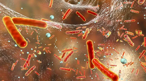
The new solution works through specially optimized deep neural networks. The cutting-edge technology works with microscopic images showing species of both bacteria and yeast-like fungi. The system analyzes these images to detect microorganisms in biological material, as well as to identify them precisely. As Dr. Bartosz Zieliński, Prof. of the Jagiellonian University, points out, one of the challenges at the stage of refining the solution was to adapt neural networks for efficient analysis based on limited input data. As a rule, neural networks work on huge amounts of data, while here the research teams had a relatively small number of clinical images and microbial culture materials for neural networks. This required conducting a series of works that adapted the specifics of neural networks for such a specific challenge.
- Our first research was relatively easy and progressed quickly. Particularly when the solution was to identify microorganisms based on photographs from cultures of well-defined bacteria or fungi. However, stairs arise when the catalog of microorganisms that the system can recognize is constantly expanding, and the microscope images show high densities of many types of bacteria. In such situations, species identification is difficult and, to the human eye, impossible. I consider refining the algorithms at this level and achieving a satisfactory prediction rate, i.e., more than 90 percent, to be our considerable achievement," adds Bartosz Zieliński, Ph.D., professor at the Jagiellonian University.
Support for traditional diagnostic methods
Importantly, based on the analysis of the image of the morphology of microorganisms, the created technology allows identification to the level of a specific species. This condition is fulfilled even when the material contains several types of bacteria within it. Currently, the system is able to identify dozens of species of bacteria and yeast-like fungi. As the developers of the solution point out, it has no limits in terms of further development and diagnostic capabilities. Ultimately, the system will recognize any species, as long as it becomes familiar with it beforehand and learns it. That's why the researchers from the Jagiellonian University, building a system library of microorganisms, started with those pathogens that are encountered most often. In the next stages, they will "teach" the system to recognize species that are less frequently found in biological materials.
According to the researchers, the system can effectively identify not only bacteria or fungi, but also protozoa, including, for example, single-celled malaria spores. The developed technology could be an interesting alternative to diagnostic method tools, which are relatively expensive to use, such as Real-time PCR, MALDI TOF, or other methods, which, it is worth noting, also do not provide immediate feedback. In addition to clinical applications, it can also work well in other areas - basically anywhere microscopic testing for detection and identification of microorganisms is needed.
- The system we have developed can ultimately serve as a stand-alone and autonomous tool for identifying microorganisms in the biological materials under study, as well as support or complement existing laboratory methods. It all depends on the model adopted in the laboratory. What is important, on the other hand, is that the test result is delivered practically immediately from the moment the image is uploaded to the system. This is a key issue in situations where the patient already has a developed infection and a quick diagnosis is needed, for example, in order to immediately implement appropriate treatment," explains Prof. Monika Brzychczy-Włoch, MD.
From the university to the clinics
UJ intends to put the new technology into clinical use as soon as possible. In order for this to happen, however, it is necessary to conduct further studies with clinical biological materials, as well as to undergo a series of procedures required for the technology to be approved for laboratory use.
- In order to pass the next stages, we need an industry partner and additional research funding. Of course, we do not exclude financing research work with public or EU funds, but it is important that the refinement of the technological capability of the method be carried out with an industry entity with relevant experience. This will definitely accelerate commercialization," assesses Gabriela Konopka-Cupial, Ph.D., Director of the Center for Technology Transfer at the Jagiellonian University, CITTRU.
The developed solution has received legal protection in the EU. As the developers emphasize, at the stage of patentability examination it was shown that this is the first technology of its kind developed in the world.